Getting rid of unwanted transformed cells: Possible new directions in cancer therapy

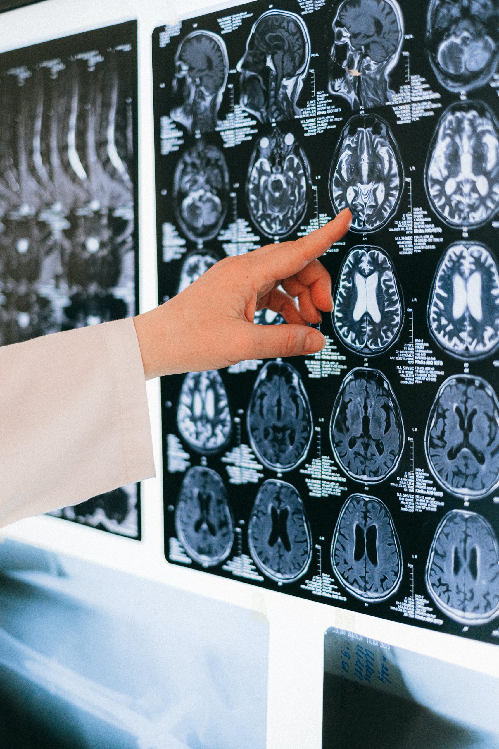
Elimination of transformed cells that can initiate cancer is necessary to maintain tissue integrity. In a new study, scientists from Tokyo University of Science show how this mechanism is regulated by the cellular process “autophagy.” They found that intact autophagic vacuoles are indispensable in mediating competitive elimination of cancer cells. Conversely, perturbation of autophagy prevents cell elimination, thereby encouraging cancer cell propagation. These findings pave the way for development of novel anti-cancer therapies.

The maintenance of a healthy cell population is a dynamic process, whereby unhealthy cells are eliminated by a defense mechanism called “cell competition”. This process is crucial as unhealthy cells or cells that have accumulated detrimental “genetic mutations” (defects in genes) over time, can initiate the formation of cancer. Cell competition is achieved by healthy normal cells that surround mutant cancer cells through various mechanisms that trigger cell removal. In addition, epithelial cells (a type of cell that constitutes external and internal body surfaces such as skin and internal organs) adopt a cell-death-independent mechanism known as “apical extrusion” to recognize and eliminate transformed cells. While the role of apical extrusion in cell competition has been well elucidated, the regulatory mechanisms underlying this complex dynamic process remain elusive.

“Autophagy” is a process by which cells degrade and recycle cellular components. Dysregulation of autophagy has been implicated in various diseases, including several cancers. While autophagy is known to facilitate the growth and survival of cancer cells at advanced stages, previous studies have indicated that autophagy may have a preventive role in early stages of cancer. Does autophagy regulate the early destruction of cancer cells through cell competition?

Building on this hypothesis, Dr. Shunsuke Kon, a junior associate professor at Tokyo University of Science along with Eilma Akter and a team of researchers, has now explored the potential regulatory role of autophagy in cell competition, in a new study recently published in Cell Reports.

Probing deeper into the possible interplay between autophagy and cell competition, the researchers used cell lines, in which cell competition is triggered by RasV12 (a cancer-causing protein). Dr. Kon explains, “We have previously shown that when a small number of mutant cells are produced in the normal epithelial layer by activating the cancer-causing gene Ras, the mutant cells are eliminated into the lumen as loser cells. This happens as a result of cell competition between the normal epithelial cells and the Ras mutant cells.”

Using the RasV12-induced mosaic (healthy + mutant cancer cells) cell competition model and fluorescent-protein labeling, the team uncovered a fascinating set of results. They showed that the RasV12-transformed cells had an increased number of autophagosomes (structures containing degradable cytoplasmic contents). Further, they noted impairment of lysosomes, the structures that fuse with autophagosomes and mediate the breakdown of their contents; which likely, caused the increase in autophagosomes. This in turn, perturbed the “autophagic flux” (a measure of autophagic degradation) in RasV12-transformed cells.

Next, they showed that the accumulated autophagosomes and the impaired lysosomes facilitated apical elimination of the transformed (cancer) cells via cell competition. These results suggest that the intact or “non-degradable” autophagosomes are important for the elimination process. Interestingly, when the researchers ablated the autophagy gene, ATG-5 in RasV12-induced cells, they noted impairment in autophagy mediated cell competition and elimination of the transformed cells. Similarly, autophagy impaired cells exhibited resistance to elimination in a mouse model, and eventually led to chronic pancreatitis or inflammation of ducts in the pancreas, thus, corroborating their earlier findings.

Together, these findings highlight the role of autophagy in competitive elimination of mutant cancer cells and tissue homeostasis (balance). The study sheds light on the role of autophagy in cancer prevention during early stages and opens avenues for the development of novel anti-cancer therapeutics.

In this context, Dr. Kon remarks, “The development of anti-cancer drugs targeting autophagy is being intensely pursued worldwide. Since the role of autophagy has been found to differ depending on the stage of cancer progression, anti-cancer strategies that take into account the stage of cancer progression can enhance treatment efficacy.”

Autophagy is surely emerging as the unsung hero that aids the removal of cancer-causing rogue cells!

***

Reference

DOI: https://doi.org/10.1016/j.celrep.2022.111292

Title of original paper: Non-degradable autophagic vacuoles are indispensable for cell competition

Journal: Cell Reports

Rates of unique names increase in Japan over 40 years from 1980s

An analysis of baby names published in municipality newsletters between 1979 and 2018 by Assistant Professor Yuji Ogihara of Tokyo University of Science and Atsuki Ito of Hitotsubashi University revealed that the rates of unique names increased in Japan over 40 years, suggesting a rise in uniqueness-seeking and individualism. This increase was observed from the 1980s, indicating that this phenomenon is not new. Their research provides important insights into changes in Japanese names and culture.

Previous research has analyzed baby names displayed by private companies and indicated that the rates of unique names increased in Japan between 2004 and 2018 (Ogihara, 2021; Ogihara et al., 2015). However, changes over a longer period were not analyzed because of the lack of a comprehensive and systematic database on baby names in Japan, unlike in other nations such as the United States and China. Therefore, it was unclear whether this increase in unique names was recent or had occurred before the 2000s. There was a possibility that the increase in unique names were found only after the 2000s.

Examining whether the rates of unique names increased for a longer period provides a betternunderstanding of not only historical changes in names and naming practices, but also cultural changes toward greater individualism which emphasizes uniqueness and independence.

To this end, Assistant Professor Yuji Ogihara of Tokyo University of Science and Atsuki Ito of Hitotsubashi University collected baby names from municipality newsletters and investigated historical changes in the rates of unique names in Japan over a longer period. Municipalities share important information such as major events (e.g., sports activities, lecture meetings), services (e.g., educational, medical), and basic statistics (e.g., financial, population) in newsletters. In these newsletters, the names of persons who are born, die, and marry in each municipality are listed.

For their study, the researchers collected municipality newsletters that fulfilled some criteria. The municipalities surveyed were geographically diverse. They were located all over Japan, from the southern part (Kyushu) to the northern part (Hokkaido). Some municipalities were located near the coast, while others were inland. The municipalities were also demographically diverse. They were located in both rural and urban areas.

The researchers analyzed 58,485 baby names published in these municipality newsletters between 1979 and 2018. They calculated the rates of the names that were not duplicated in each of the municipalities in each year. Then, they analyzed their historical changes. Furthermore, they calculated the rates of unique names not only within a given year (e.g., 2000) but also within a three-year unit (the target year, the year before it, and the year after it; e.g., 1999, 2000, 2001), and performed the same analysis.

They found that the rates of unique names increased within both time frames. Thus, unique names increased not only after the 2000s, but also from the 1980s for 40 years. This result shows that parents increasingly gave unique names to their babies and that Japanese culture increasingly emphasized uniqueness and independence for the 40 years, providing further evidence of the rise in uniqueness-seeking and individualism in Japan. This finding is also consistent with prior studies showing the rise in individualism in other aspects such as family structure and values.

Moreover, the findings reported in previous research (Ogihara, 2021; Ogihara et al., 2015) were replicated in this study: unique names increased in Japan in the 2000s and 2010s. In addition, the rates of unique names increased more rapidly for girls than for boys. This result may suggest that parents came to have stronger hope for their daughters to become unique and independent than for their sons. This means that the same phenomena were observed in a dataset different from that analyzed in previous research, indicating that the finding of an increase in unique names in Japan is robust. These findings were made available online on April 28, 2022, and published on June 21, 2022, in volume 3 of the international journal Current Research in Ecological and Social Psychology.

This study reveals an increase in the rates of unique names in Japan via an analysis of baby names published in municipality newsletters. Unique names increased from at least the 1980s in Japan. It shows that Japanese culture has changed toward greater individualism which emphasizes uniqueness and independence. Therefore, this research contributes to the understanding of changes in not only Japanese names and naming practices but also Japanese culture.

Assistant Professor Ogihara plans to continue investigating the historical changes in names and naming practices in Japan. In the near future, he aims to examine whether these changes have continued in the last few years recently and how COVID-19 has affected naming practices in Japan.

Reference

DOI: https://doi.org/10.1016/j.cresp.2022.100046

New magnesium superionic conductor towards lithium-free solid-state batteries

As we move towards a more energy-efficient society, the need for high-capacity, cost-effective batteries is greater than ever. Magnesium is a promising material for such solid-state batteries owing to its abundance, but its practical application is limited by the poor conductivity of magnesium ions (Mg2+) in solids at room temperature. Recently, researchers from Japan have developed a novel Mg2+ conductor with a practically applicable superconductivity of 10-3 S cm-1, overcoming this decades-long roadblock.

The development of highly efficient energy storage devices that can store renewable energy is crucial to a sustainable future. In today’s world, solid-state rechargeable lithium ion (Li+) batteries are the state of the art. But lithium is a rare earth metal, and society’s dependence on the element is likely to lead to a rapid decline in resources and subsequent price hikes.

Magnesium ion (Mg2+)-based batteries have gained momentum as an alternative to Li+. The earth’s crust holds ample magnesium, and Mg2+-based energy devices are said to have high energy densities, high safety, and low cost. But the wide application of Mg2+ is limited by its poor conductivity in solids at room temperature. Mg2+ has poor solid-state conductivity because divalent positive ions (2+) experience strong interactions with their neighboring negative ions in a solid crystal, impeding their migration through the material.

This hurdle was recently overcome by a research team from the Tokyo University of Science (TUS). In their new study published online on 4 May 2022 and on 18 May 2022 in volume 144 issue 19 of the Journal of the American Chemical Society, they report for the first time, a solid-state Mg2+ conductor with superionic conductivity of 10−3 S cm−1 (the threshold for practical application in solid-state batteries). This magnitude of conductivity for Mg2+ conductors is the highest reported to date. According to Junior Associate Professor Masaaki Sadakiyo of TUS, who led the study, “In this work, we exploited a class of materials called metal–organic frameworks (MOFs). MOFs have highly porous crystal structures, which provide the space for efficient migration of the included ions.

Here, we additionally introduced a “guest molecule,” acetonitrile, into the pores of the MOF, which succeeded in strongly accelerating the conductivity of Mg2+.” The research group further included Mr. Yuto Yoshida, also from TUS, Professor Teppei Yamada from The University of Tokyo, and Assistant Professor Takashi Toyao and Professor Ken-ichi Shimizu from Hokkaido University. The paper was made available online on May 4, 2022 and was published in Volume 144 Issue 19 of the journal on May 18, 2022.

The team used a MOF known as MIL-101 as the main framework and then encapsulated Mg2+ ions in its nanopores. In the resultant MOF-based electrolyte, Mg2+ was loosely packed, thereby allowing the migration of divalent Mg2+ ions. To further enhance ion conductivity, the research team exposed the electrolyte to acetonitrile vapors, which were adsorbed by the MOF as guest molecules.

The team then subjected the prepared samples to an alternating current (AC) impedance test to measure ionic conductivity. They found that the Mg2+ electrolyte exhibited a superionic conductivity of 1.9 × 10⁻³ S cm−1. This is the highest ever reported conductivity for a crystalline solid containing Mg2+.

To understand the mechanism behind this high conductivity, the researchers carried out infrared spectroscopic and adsorption isotherm measurements on the electrolyte. The tests revealed that the acetonitrile molecules adsorbed in the framework allowed for the efficient migration of the Mg2+ ions through the body of the solid electrolyte.

These findings of this study not only reveal the novel MOF-based Mg2+ conductor as a suitable material for battery applications, but also provide critical insights into the development of future solid-state batteries. “For a long time, people have believed that divalent or higher valency ions cannot be efficiently transferred through a solid. In this study, we have demonstrated that if the crystal structure and surrounding environment are well-designed, then a solid-state high-conductivity conductor is well within research,” explains Dr. Sadakiyo.

When asked about the research group’s future plans, he reveals, “We hope to further contribute to society by developing a divalent conductor with even higher ionic conductivity.”

We look forward to seeing what they develop next!

Mechanisms driving 1,2-Dichloropropane-induced cancer in printing industry

Study finds that when exposed to 1,2-dichloropropane, cells show altered gene expression that induces cellular cascades promoting cancer

1,2-dichloropropane (1,2-DCP) is a solvent used in the printing industry. It was linked to cholangiocarcinoma in 2013, when printing company employees exposed to 1,2-DCP were diagnosed with the cancer. To understand the genes influencing cholangiocarcinoma development, scientists examined gene expression profiles in co-cultured cholangiocytes and macrophages exposed to 1,2-DCP. They found DNA repair genes in cholangiocytes and cell cycle genes in macrophages were upregulated, yielding novel insights on the pathogenesis of this elusive occupational hazard.

1,2-Dichloropropane (1,2-DCP) is a solvent widely used in the printing industry. It rose to prominence when it was linked to the development of cholangiocarcinoma, or bile duct cancer, in the employees of an offset printing firm in Osaka in 2013. Thereafter, the International Agency for the Research on Cancer reclassified 1,2-DCP as being carcinogenic to humans, and many studies have since focused on occupational cholangiocarcinoma on exposure to 1,2-DCP.

Common cholangiocarcinoma develops in the cholangiocytes (or epithelial cells) of the bile duct and liver. On the other hand, occupational cholangiocarcinoma has markedly different features, such as the presence of non-characteristic precancerous lesions and inflammatory changes in the surrounding non-cancerous tissue. Research suggests that while 1,2-DCP primarily targets cholangiocytes, it indirectly damages their DNA in the presence of inflammatory cells called macrophages. However, the exact mechanism of 1,2-DCP-induced cholangiocarcinoma remains a mystery.

To solve this problem, in a new study, a group of researchers led by Professor Gaku Ichihara from Tokyo University of Science (TUS), identified the gene expression profiles of cholangiocytes co-cultured with macrophages and exposed to 1,2-DCP. Prof. Ichihara says, “Our findings identified the upregulation of genes tied to DNA repair and the cell cycle in cholangiocytes and macrophages, respectively. This suggests that the DNA damage, cell proliferation, and ultimately neoplasia occurring in the bile ducts is likely driven by the altered cell function induced by the abnormal gene expression.”

In the study, published in the journal Scientific Reports (published online on 02 July 2022), Prof. Ichihara, together with his colleagues Shigeyuki Shichino and Kouji Matsushimia at TUS, Kazuo Kinoshita from Shizuoka Graduate University of Public Health, and Sahoko Ichihara from Jichi Medical University School of Medicine, co-cultured cholangiocytes and macrophages that were exposed to varying concentrations of 1,2-DCP for 24 hours. The concentrations selected mirrored the occupational exposure of workers in a poorly ventilated environment.

Prof. Ichihara’s previous work had shown that in the presence of macrophages, 1,2-DCP induced the expression of activation-induced cytidine deaminase, which is a DNA-mutating enzyme, along with excess DNA damage and reactive oxygen species production in cholangiocytes. To delve deeper, the team used transcriptomics to study the gene expression patterns in the cells and identify the intracellular mechanisms driving carcinoma formation.

The data revealed that in the presence of 1,2-DCP, co-cultured cholangiocytes showed higher expression of base excision repair genes, whereas macrophages revealed upregulation of cell cycle genes. “The upregulation of DNA repair genes suggests an increase in DNA damage as 1,2-DCP concentration increases. Furthermore, macrophages could proliferate at a given site following 1,2-DCP exposure. Since they play an important role in the regulation of inflammatory responses by releasing cytokines and signaling molecules, their overstimulation could result in the persistent production of these compounds which ultimately influence various pathological states and cancer,” explains Prof. Ichihara.

The implications of the study are far-reaching in the fields of environmental toxicology and occupational cancer prevention. The team’s findings show that it is possible to pinpoint how potential carcinogens promote cancer without directly damaging DNA. Prof. Ichihara and his team are confident they can build on their findings and design further studies to fully understand the cross talk between cholangiocytes and macrophages and elucidate the mechanisms behind the erroneous DNA damage repair in cholangiocytes.

***

Reference

Title of original paper: Transcriptome analysis of human cholangiocytes exposed to carcinogenic 1,2‑dichloropropane in the presence of macrophages in vitro

Journal: Scientific Reports

DOI: https://doi.org/10.1038/s41598-022-15295-3


Changes in protein structure, assembly with fluoride nanoparticles and coexisting ions

Protein function and activity is determined by both their assembly and secondary structure. Abnormalities related to either protein aggregation or secondary structure can lead to neurodegenerative diseases. In a new study, an international research team reveal how fluoride nanoparticles, materials used in in vivo imaging, affect the assembly and structure of the amyloid β protein. Their results present a step towards better treatment and prevention of neurologic disorders like Alzheimer’s disease.

Self-assembly, or the association of individual units of a material into ordered structures or patterns, is a phenomenon of great research interest for materials scientists. One prominent example of self-assembly comes from the self-assembly of proteins in biological systems. The function and activity of proteins are governed by their assembly state. Additionally, the protein’s “secondary structure,” characterized by its folding into structures, such as a β-sheet, also plays a role. In fact, abnormalities in the protein secondary structures or their assembly can lead to various neurodegenerative diseases, including Alzheimer’s disease.

Nanoparticles (NPs) offer a promising route for the treatment and prevention of such diseases by allowing a controlled and targeted drug delivery. Additionally, inorganic NPs, such as fluoride NPs, are used in brain imaging applications. Compared to organic NPs, inorganic NPs are considered a better candidate for developing high functional materials. But, there is much concern regarding their bio-toxicity. While their interactions with bioproteins have been studied, the mechanism underlying these interactions are not well understood.

An international team of scientists from Tokyo University of Science (TUS) in Japan and Nazarbayev University in Kazakhstan has now addressed this issue. In their study, which was made available online on June 2, 2022, and was published in Volume 5, Issue 6 the journal ACS Applied Bio Materials on June 20, 2022, the team investigated a section of the amyloid β peptide (a protein found in the plaques forming in the brains of patients with Alzheimer’s disease) in solution with fluoride ceramic (CeF3) NPs. The study was led by Junior Associate Professor Masakazu Umezawa and included contributions from Mr. Naoya Sakaguchi from TUS and Assistant Professors Mehdi Amouei Torkmahalleh and Dhawal Shah from Nazarbayev University.

The team used a technique called “Fourier transform infrared spectroscopy” (FTIR) to directly monitor the effect of the NP surface on the peptide bonds. “We found that, near the nanoparticle surface, peptides are more likely to form β-sheets. This comes as an effect of hydrophobicity. The parts of the peptide that repelled by the water solution stick to the nanoparticles, and form aggregates more easily,” explains Dr. Umezawa.

In addition, the team investigated the effect of other surrounding ions in the solution. “What we found was very surprising. Even without the nanoparticles, the environment affected the rate of secondary structure formation,” says Dr. Umezawa, “This effect, resulting from a combination of electrostatic interaction and hydrogen bonding, was exaggerated upon adding nanoparticles. With a careful choice of ions and nanoparticles, the β-sheet formation can be either suppressed or promoted. This implies that the process can be controlled and engineered to eradicate adverse effects.”

The experimental results were complemented with molecular dynamics simulations performed by the Nazarbayev University team. This, in turn, helped design and guide the experiments as well as provide insights into the results.

With this deeper understanding of the interaction between proteins and NPs, the study paves the way for controlled protein folding processes. With such control, any protein deformations could be eliminated, and positive interactions and structural changes could be promoted. This could lead to a better prevention and treatment protocol for Alzheimer’s disease and, eventually, to a better quality of life for aged adults.

***

Reference

Title of original paper: Changes in the Secondary Structure and Assembly of Proteins on Fluoride Ceramic (CeF3) Nanoparticle Surfaces

Journal: ACS Applied Bio Materials

DOI: https://doi.org/10.1021/acsabm.2c00239

Scientists uncover new therapeutic target for treating colorectal tumours

Colorectal tumors are a common adverse effect of chronic inflammation of the intestine. In a new study, researchers from Japan and China demonstrate that mice models with dendritic cell immunoreceptor (DCIR) protein deficiency are resistant to colonic tumors induced by dextran sodium sulfate and azoxymethane. Further, they identify an antibody that reduces colitis severity and colonic tumor growth, highlighting the potential of DCIR as a therapeutic target.

Inflammatory bowel disease (IBD) is an umbrella term for two diseases, Crohn’s disease and ulcerative colitis, that are characterized by the prolonged inflammation of the gastrointestinal tract. This condition often leads to the development of colorectal tumors. Understanding the pathogenesis of IBD is, therefore, crucial to mitigate the incidence of colonic tumors.

It turns out that innate immune receptors, particularly those expressed in the gut, such as C-type lectin receptors (CLRs), are responsible for the development of IBD. However, CLRs also play a vital role in the regulation of gut microbiota and defense against pathogens. As a result, a balance needs to be struck to maintain intestinal homeostasis.

Dendritic cell immunoreceptor (DCIR) is one such CLR that is responsible for maintaining homeostasis of the immune and skeletal systems. Previous studies have suggested that DCIR negatively regulates both innate and acquired immune responses. Blocking DCIR could, therefore, potentially boost immunity against colon tumor. However, its role in intestinal immunity has remained unclear.

Against this backdrop, a research group led by Professor Yoichiro Iwakura of Tokyo University of Science (TUS) in Japan has now shed light on this issue. In their study, to be published online on 02 August 2022 in the international journal Cell Reports, the group studied the development of colitis and colon tumor in mice models deficient in DCIR.

To this end, the group fed the mice with drinking water containing dextran sodium sulfate (DSS), a synthetic sulfated polysaccharide, and azoxymethane (AOM), a neurotoxic chemical, to induce colon tumors similar to that observed in humans with IBD.

To their surprise, they found that the mice lacking DCIR showed reduced colitis severity and AOM-DSS–induced colorectal tumor growth. Moreover, compared to the wild-type mice (control), the DCIR-deficient mice showed lower body weight loss as well as reduced proinflammatory cell infiltration in the colon.

What do these observations imply? Prof. Iwakura explains, “Our findings point to the fact that intestinal carcinogenesis and inflammation are facilitated by DCIR signalling, which points to the possibility that blocking DCIR might prevent ulcerative colitis and colon cancer.”

Corroborating this possibility, the study further revealed that the use of an antibody called “anti-NA2” against asialo-biantennary-N-glycans (NA2), a ligand (binding molecule) to DCIR, reduced DSS colitis symptoms and prevented colorectal tumor growth.

The researchers are excited by these findings. Speaking about the practical applications of their study, Prof. Iwakura says, “Our results suggest that therapeutics targeting DCIR and its ligands could be used to effectively treat autoimmune diseases, IBD, and cancer, which have been traditionally difficult to treat.”

Sure enough, this study could open doors to novel therapeutic strategies for treating colorectal tumors, improving not only the lives of patients with IBD but also our understanding of the pathogenesis of human diseases.

***

Reference

Title of original paper: Blocking DCIR mitigates colitis and prevents colorectal tumors by enhancing the GM-CSF-STAT5 pathway

Journal: Cell Reports

DOI: https://doi.org/10.1016/j.celrep.2022.111158

About The Tokyo University of Science

Tokyo University of Science (TUS) is a well-known and respected university, and the largest science-specialized private research university in Japan, with four campuses in central Tokyo and its suburbs and in Hokkaido. Established in 1881, the university has continually contributed to Japan’s development in science through inculcating the love for science in researchers, technicians, and educators.

With a mission of “Creating science and technology for the harmonious development of nature, human beings, and society”, TUS has undertaken a wide range of research from basic to applied science. TUS has embraced a multidisciplinary approach to research and undertaken intensive study in some of today’s most vital fields. TUS is a meritocracy where the best in science is recognized and nurtured. It is the only private university in Japan that has produced a Nobel Prize winner and the only private university in Asia to produce Nobel Prize winners within the natural sciences field.

Website: https://www.tus.ac.jp/en/mediarelations/

About Professor Yoichiro Iwakura from Tokyo University of Science

Professor Yoichiro Iwakura has been the director of the Center for Animal Disease Models at the Tokyo University of Science (TUS) since 2013, and has published numerous papers since his graduation from Kyoto University in 1970. He started researching interferon proteins at the university’s Institute for Virus Research before moving to the Sloan-Kettering Cancer Institute in the United States to analyze the developmental mechanism of early mouse embryos. In 1985, he moved to the University of Tokyo where he generated more than 100 lines of gene-modified mice as the director of the Center for Experimental Medicine to analyze the pathogenesis of infectious and autoimmune diseases.

He retired from the University of Tokyo and became an Emeritus Professor in 2012. He then moved to TUS. He has been a visiting professor for many universities, including Dalian Medical University in China and Chiba University in Japan. His work involves the development and analysis of animal disease models and research on autoimmune diseases and infectious diseases. He won the Hideyo Noguchi Memorial Award for Medical Science in 2015 and was selected as a highly cited researcher (Thomson Reuters and Clarivate Analytics) for six consecutive years from 2014.

Investigating the role of policy disagreements in party switching using a Japanese example

In 2017, the Japanese party system took a dramatic turn when the major opposition party split, turning the election into a three-way contest. Long-dormant policy disagreements prompted legislators to switch parties. To understand why, researchers from Japan have recently analyzed post-electoral expert survey data. Their findings reveal both the reasons for the separation and the basis for party cohesion, offering valuable insights into party unity and cohesion.

The transformation of party politics in Japan has been marked by an array of sensational events, from the dominance of the Liberal Democratic Party (LDP) to party fragmentation. Individual legislators switch parties for a number of reasons, including policy considerations. The payoffs from “party switching” are closely linked to electoral and party systems, leading to a shift in the dynamics of party competition.

In Japan, such a situation of party switching was witnessed before the 2017 election. The founding leader of a new party, “Hope,” aggressively politicized an inactive debate regarding defense policy. Several members were persuaded to join the Hope, resulting in the collapse of the largest opposition group, “Minshin,” into three parties. However, there remains a lack of explanation as to why the long-dormant disagreement culminated in such a severe rupture.

To uncover the underlying rationale, a Japanese research group consisting of Junior Associate Professor Tomoko Matsumoto of Tokyo University of Science, Associate Professor Hiroki Kubo of Meiji Gakuin University, and Professor Kentaro Yamamoto of Hokkai-Gakuen University, examined responses on “issue position” and the “salience of party policies” from an expert survey conducted shortly after the 2017 general elections. Speaking of the motivation of the study, Dr. Matsumoto says, “The frequent breakdowns of opposition parties have created uncertainty among voters. By analyzing the expert survey, we hope to understand the structure of oppositional party policies in Japan.” Their seminal study was recently published in Japanese Journal of Political Studies.

The researchers began by assessing the expert responses using the “differential-item functioning (DIF)” analysis of “Aldrich-McKelvey” scaling. They next employed “Blackbox transpose” scaling to examine the multidimensional nature of the issue stances. One important point raised by this study is that traditional DIF analysis assumes diversity in responses is due to perceptional limitations and biases. However, the diversity in this case meant that the parties were unable to discern and convey their policy positions. As a result, a corrected version of the DIF analysis is required. “When we performed the corrected DIF analysis, we realized that the corrected value differed from the average value of the raw data obtained from the responses. In other words, the importance of using responses after rectification was reaffirmed,” reveals Dr. Matsumoto. “This is a huge revelation since online surveys are increasingly being used for scientific research.”

The results revealed a strong discord among the splinter parties over defense policy. However, their opinions on the environment, decentralization, and other policies clearly coincided. This was most likely the foundation for the main opposition camp’s unity. Furthermore, DIF analysis revealed considerable differences in expert responses on the relevance of defense policy. As a result, the rapid politicization of defense policy may have contributed to its relevance, which in turn led to the split.

Although the study showed that the major opposition separated over defense policy disagreements, it also showed that splinter opposition parties agreed on environmental and decentralization policies. Dr. Matsumoto concludes, “Our findings suggest that party switching is triggered not just by policy disagreement, but also by the decision of which policy is politicized. This may help us comprehend the future dynamics between the ruling and opposition parties.”

Party competition and party systems are critical facets of parliamentary democracies. These findings go a long way towards improving our understanding of these political structures.

***

Reference

Title of original paper: Party Switching and Policy Disagreement: Scaling Analysis of Experts’ Judgement

Journal: Japanese Journal of Political Studies

DOI: https://doi.org/10.1017/S1468109922000160

Ultrasound exposure improves depressive behavior in rodents: Study

It is well known that whole-body exposure to high frequency ultrasound increases brain activity in humans. However, little is known about its impact and associated mechanisms on emotional states like depression. Now, a team of researchers at Tokyo University of Science have recently demonstrated the anti-depressant effects of ultrasound exposure in a rodent model of depression. Their findings shed light on the potential of ultrasound exposure as a non-invasive treatment for mental disorders.

The effect of ultrasound waves on the function of the human brain has been the key focus of recent research, which has indicated its potential as an effective, non-invasive approach for the modulation of brain activity. While the effects of ultrasound exposure on consciousness and cognition have been extensively explored, little is known about its impact on emotional states such as depression. To add to it, there are limitations in our understanding of neural and molecular mechanisms that underpin emotions.

Fortunately, rats experience pleasant emotions in response to high-frequency ultrasound vocalizations (USVs), making them ideal model organisms to study mechanisms underlying depression.

To this end, a team of researchers led by Professor Akiyoshi Saitoh, including Professor Satoru Miyazaki, Assistant Professor Daisuke Yamada and Ms. Tsugumi Yamauchi from Tokyo University of Science, and Mr. Shoichi Nishino from FUJIMIC, Inc., delved deeper into understanding the effects of ultrasound exposure on depression, by conducting experiments on rats lacking olfactory lobes—organs that regulate neurotransmission. These “olfactory bulbectomized (OB)” rats undergo changes in neurotransmitters, endocrine secretions, and behavior, which are similar to those observed in humans with depression.

Giving further insights into their study, Prof. Saitoh remarked, “Since studies on ultrasound exposure have been primarily conducted on human subjects, we needed to establish robust animal models to elucidate underlying mechanisms using invasive techniques. In our current study, we have used OB rats to study the effects of ultrasound on neural activity and behavior” Their study, published in Volume 33, Issue 10 of NeuroReport on July 6, 2022, is the first of its kind to demonstrate potential anti-depressant effects of ultrasound exposure in rats.

Initially, the team exposed wild type and OB rats to USV for 24 hours, following which they scored them for “hyperemotionality” (agitation and anxiety-like behavior) by studying their responses to getting attacked, getting startled, facing a struggle, and initiating a fight.

Next, they monitored plasma corticosterone (a hormone that is released in response to stress) levels in the blood samples of these rats. In addition, the team assessed anxiety-like behavior of the rodents using the elevated plus maze (EPM)—an approach which triggers behavioral anxiety in rats by exposing them to open spaces in a maze, and causes them to move to closed spaces.

Their findings revealed that OB rats exposed to USV had significantly lower hyperemotionality scores and lower plasma corticosterone levels than unexposed rats. Furthermore, in OB rats with a higher latency initially. i.e., higher inclination to reach the open areas of the maze, ultrasound exposure significantly decreased their latency. Similar effects were observed with a 50-kHz ultrasound frequency which was generated artificially.

This study provides novel evidence on the anti-depressant effects of ultrasound exposure in rodents. “Our findings suggest that OB rats may be a useful animal model for investigating the effects of ultrasound exposure and mechanisms of influence.”, exclaims Prof. Saitoh about the implications of the study.

He further adds, “Unlike drug therapy, ultrasound exposure is non-invasive and easy to use. An ultrasound based therapeutic device may therefore aid the treatment and prevention of mental disorders in patients while they go about their daily lives.”

Let’s hope that these results pave the way for developing ultrasound exposure therapy as a novel treatment to help patients cope with stress and psychiatric disorders.

***

Reference

Title of original paper: High-frequency ultrasound exposure improves depressive-like behavior in an olfactory bulbectomized rat model of depression

Journal: NeuroReport

DOI: https://doi.org/10.1097/WNR.0000000000001804

Self-assembling 2D materials at a liquid–liquid interface

Molecular 2D materials find immense applications in materials science, owing to their wide structural variety and easy controllability. Establishing a simple and efficient method for their synthesis is, therefore, important. Now, scientists from Japan present a simple method for synthesizing heterolayer coordination nanosheets, a promising 2D material, shedding light on how certain chemical coordination reactions occur at liquid–liquid interfaces. Their method could help develop novel 2D materials with applications in optoelectronic devices.

The past few decades have witnessed a great amount of research in the field of two-dimensional (2D) materials. As the name implies, these thin film-like materials are composed of layers that are only a few atoms thick. Many of the chemical and physical properties of 2D materials can be fine-tuned, leading to promising applications in many fields, including optoelectronics, catalysis, renewable energy, and more.

Coordination nanosheets are one particularly interesting type of 2D material. The “coordination” refers to the effect of metallic ions in these molecules, which act as coordination centers. These centers can spontaneously create organized molecular dispositions that span multiple layers in 2D materials. This has attracted the attention of materials scientists due to their favorable properties. In fact, we have only begun to scratch the surface regarding what heterolayer coordination nanosheets – coordination nanosheets whose layers have different atomic composition – can offer.

In a recent study published first on June 13, 2022, and featured on the front cover of Chemistry—A European Journal, a team of scientists from Tokyo University of Science (TUS) and The University of Tokyo in Japan reported a remarkably simple way to synthesize heterolayer coordination nanosheets. Composed of the organic ligand, terpyridine, coordinating iron and cobalt, these nanosheets assemble themselves at the interface between two immiscible liquids in a peculiar way. The study, led by Prof. Hiroshi Nishihara from TUS, also included contributions from Mr. Joe Komeda, Dr. Kenji Takada, Dr. Hiroaki Maeda, and Dr. Naoya Fukui from TUS.

To synthesize the heterolayer coordination nanosheets, the team first created the liquid–liquid interface to enable their assembly. They dissolved tris(terpyridine) ligand in dichloromethane (CH2Cl2), an organic liquid that does not mix with water. They then poured a solution of water and ferrous tetrafluoroborate, an iron-containing chemical, on top of the CH2Cl2. After 24 hours, the first layer of the coordination nanosheet, bis(terpyridine)iron (or “Fe-tpy”), formed at the interface between both liquids.

Following this, they removed the iron-containing water and replaced it with cobalt-containing water. In the next few days, a bis(terpyridine)cobalt (or “Co-tpy”) layer formed right below the iron-containing one at the liquid–liquid interface.

The team made detailed observations of the heterolayer using various advanced techniques, such as scanning electron microscopy, X-ray photoelectron spectroscopy, atomic force microscopy, and scanning transmission electron microscopy. They found that the Co-tpy layer formed neatly below the Fe-tpy layer at the liquid–liquid interface. Moreover, they could control the thickness of the second layer depending on how long they left the synthesis process run its course.

Interestingly, the team also found that the ordering of the layers could be swapped by simply changing the order of the synthesis steps. In other words, if they first added a cobalt-containing solution and then replaced it with an iron-containing solution, the synthesized heterolayer would have cobalt coordination centers on the top layer and iron coordination centers on the bottom layer. “Our findings indicate that metal ions can go through the first layer from the aqueous phase to the CH2Cl2 phase to react with terpyridine ligands right at the boundary between the nanosheet and the CH2Cl2 phase,” explains Prof. Nishihara. “This is the first ever clarification of the growth direction of coordination nanosheets at a liquid/liquid interface.”

Additionally, the team investigated the reduction–oxidation properties of their coordination nanosheets as well as their electrical rectification characteristics. They found that the heterolayers behaved much like a diode in a way that is consistent with the electronic energy levels of Co-tpy and Fe-tpy. These insights, coupled with the easy synthesis procedure developed by the team, could help in the design of heterolayer nanosheets made of other materials and tailored for specific electronics applications. “Our synthetic method could be applicable to other coordination polymers synthesized at liquid–liquid interfaces,” highlights Prof. Nishihara. “Therefore, the results of this study will expand the structural and functional diversity of molecular 2D materials.”

With eyes set on the future, the team will keep investigating chemical phenomena occurring at liquid–liquid interfaces, elucidating the mechanisms of mass transport and chemical reactions. Their findings can help expand the design of 2D materials and, hopefully, lead to better performance of optoelectronic devices, such as solar cells.

***

Reference

Title of original paper: Chemically Laminated 2D Bis(terpyridine)metal Polymer Films: Formation Mechanism at the Liquid–Liquid Interface and Redox Rectification

Journal: Chemistry—A European Journal

DOI: https://doi.org/10.1002/chem.202201316

Scientists discover mechanism behind suppression of fearful memories

Fearful events negatively impact the brain.

For instance, war veterans often go through post-traumatic stress disorder months after the cessation of the triggering event. Now, in a study led by Tokyo University of Science researchers, the precise mechanism of suppression of such fearful memories has been uncovered. Using a mouse model, the researchers identified the associated biochemical pathways, thus paving the way for the development and clinical evaluation of therapeutic compounds such as KNT-127.

Tragic events like wars, famines, earthquakes, and accidents create fearful memories in our brain. These memories continue to haunt us even after the actual event has passed. Luckily, researchers from Tokyo University of Science (TUS) have recently been able to understand the hidden biochemical mechanisms involved in the selective suppression of fearful memories, which is called fear extinction.

The researchers, who had previously demonstrated fear extinction in mice using the chemically synthesized compound “KNT-127,” have now identified the underlying mechanism of this compound’s action. Their findings have been published recently in Frontiers in Behavioral Neuroscience.

Prof. Akiyoshi Saitoh, lead author of the study, and Professor at TUS, muses, “Drugs that treat fear-related diseases like anxiety and posttraumatic stress disorder must be able to help extinguish fear. We previously reported that KNT-127, a selective agonist of the d-opioid receptor or DOP, facilitates contextual fear extinction in mice. However, its site of action in the brain and the underlying molecular mechanism remained elusive. We therefore investigated brain regions and cellular signaling pathways that we assumed would mediate the action of KNT-127 on fear extinction.”

“We investigated the molecular mechanism of KNT-127-mediated suppression of fearful memories. We administered KNT-127 to specific brain regions and identified the brain regions involved in promoting fear extinction via delta receptor activation,” elaborates Dr. Daisuke Yamada, co-author of the study, and Assistant Professor at TUS.

Using a mouse model, the research team performed fear conditioning test on laboratory mice. During fear conditioning, mice learn to associate a particular neutral conditioned stimulus with an aversive unconditioned stimulus (e.g., a mild electrical shock to the foot) and show a conditioned fear response (e.g., freezing).

After the initial fear conditioning, the mice were re-exposed to the conditioning chamber for six minutes as part of the extinction training. Meanwhile, the fear-suppressing therapeutic “KNT-127” was microinjected into various regions of the brain, 30 minutes prior to re-exposure. The treated brain regions included the basolateral nucleus of the amygdala (BLA), the hippocampus (HPC), and the prelimbic (PL) or infralimbic subregions (IL) of the medial prefrontal cortex. The following day, the treated mice were re-exposed to the chamber for six minutes for memory testing.

The fear-suppressing “KNT-127” that infused into the BLA and IL, but not HPC or PL, significantly reduced the freezing response during re-exposure. Such an effect was not observed in mice that did not receive the KNT-127 treatment, thus confirming the fear-suppressing potential of this novel compound.

Chemical compounds known to inhibit the actions of key intracellular signaling pathways like PI3K/Akt and MEK/ERK pathways reversed the therapeutic effect, thereby suggesting the key roles of these two pathways in influencing KNT-127-mediated fear extinction.

The first author of the study, Ayako Kawaminami, who is currently pursuing research at TUS, says, “The selective DOP antagonist that we used for pretreatment antagonized the effect of KNT-127 administered into the BLA and IL. Further, local administration of MEK/ERK inhibitor into the BLA and of PI3K/Akt inhibitor into the IL abolished the effect of KNT-127. These findings strongly indicated that the effect of KNT-127 is mediated by MEK/ERK signaling in the BLA, by PI3K/Akt signaling in the IL, and by DOPs in both brain regions. We have managed to show that DOPs play a role in fear extinction via distinct signaling pathways in the BLA and IL.”

PTSD and phobias are thought to be caused by the inappropriate or inadequate control of fear memories. Currently, serotonin reuptake inhibitors and benzodiazepines are prescribed during therapy. However, many patients do not derive significant therapeutic benefits from these drugs. Therefore, there is an urgent need for the development of new therapeutic agents that have a different mechanism of action from existing drugs.

Dr. Hiroshi Nagase, a Professor at University of Tsukuba and a coauthor of the study, concludes, “We have succeeded in creating KNT-127 by successfully separating convulsion- and catalepsy-inducing actions, which has so far been extremely difficult. Our findings will provide useful and important information for the development of evidence-based therapeutics with a new mechanism of action, that is targeting DOP.”

Fighting fear with the right therapeutic is the need of the hour, as anxiety and stress increase globally, and the findings of this study could help us achieve this objective. We have our fingers crossed.

***

Reference

Title of original paper: Selective δ-Opioid Receptor Agonist, KNT-127, Facilitates Contextual Fear Extinction via Infralimbic Cortex and Amygdala in Mice

Journal: Frontiers in Behavioral Neuroscience

DOI: https://doi.org/10.3389/fnbeh.2022.808232

About The Tokyo University of Science

Tokyo University of Science (TUS) is a well-known and respected university, and the largest science-specialized private research university in Japan, with four campuses in central Tokyo and its suburbs and in Hokkaido. Established in 1881, the university has continually contributed to Japan’s development in science through inculcating the love for science in researchers, technicians, and educators.

With a mission of “Creating science and technology for the harmonious development of nature, human beings, and society”, TUS has undertaken a wide range of research from basic to applied science. TUS has embraced a multidisciplinary approach to research and undertaken intensive study in some of today’s most vital fields. TUS is a meritocracy where the best in science is recognized and nurtured. It is the only private university in Japan that has produced a Nobel Prize winner and the only private university in Asia to produce Nobel Prize winners within the natural sciences field.

Website: https://www.tus.ac.jp/en/mediarelations/

About Professor Akiyoshi Saitoh from Tokyo University of Science

Dr. Akiyoshi Saitoh is serving as a Professor in the Department of Pharmacy, at the Tokyo University of Science, Japan. His research work primarily focuses on the role of the amygdala in the rodent fear extinction memory as well as on the development of novel opioid delta receptor agonists for combating depression and anxiety. Prof. Saitoh has published over 100 refereed papers so far. He also has a patent to his credit.